Worksheet Design and Functionality

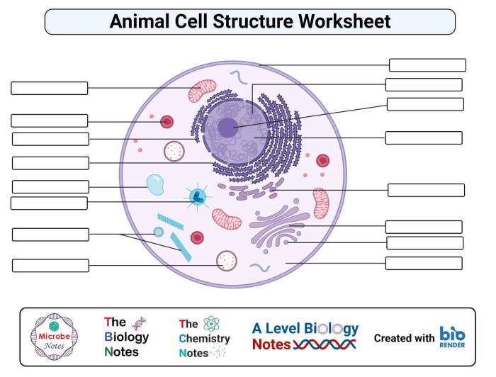
Animal cell coloring labeling worksheet – This section details the design and functionality of a coloring worksheet aimed at reinforcing understanding of animal cell structure. The worksheet employs a simplified diagram to facilitate ease of use and comprehension, particularly for younger learners. The design prioritizes clarity and visual appeal to engage students actively in the learning process.This worksheet uses a visually appealing approach to learning about animal cell components.
The simplified diagram makes it accessible for various age groups and learning styles, while the coloring activity enhances engagement and retention. The accompanying labeling exercise strengthens understanding of the function and location of each organelle.
Simplified Animal Cell Diagram
The worksheet features a large, central illustration of an animal cell. This diagram is simplified to show only the essential organelles, avoiding unnecessary detail that might confuse beginners. The organelles included are: the nucleus (large, centrally located, colored a light purple), the cell membrane (thin outer boundary, light blue), the cytoplasm (filling the cell, light yellow), the mitochondria (oval-shaped, scattered throughout the cytoplasm, dark orange), the ribosomes (small dots scattered in the cytoplasm, dark red), the endoplasmic reticulum (network of interconnected membranes, light green), the Golgi apparatus (stack of flattened sacs, light brown), and the lysosomes (small, circular organelles, dark purple).
Understanding animal cell structures is made engaging with an animal cell coloring labeling worksheet. This activity helps visualize the intricate components within. Interestingly, the concept of camouflage, as seen in a fantastic resource like this animal camouflage coloring page , highlights how animals adapt to their environments, a concept that can be further explored by relating it back to the functions of different organelles within the animal cell on your worksheet.
Returning to the worksheet, accurate labeling is key to mastering the complexities of animal cell biology.
Each organelle is clearly delineated and labeled with a numbered key for easy identification. The overall visual design is clean and uncluttered, with clear lines and easily distinguishable colors.
Instructions for Completing the Worksheet
The following instructions are provided to guide students through the worksheet activity:
- Carefully color each organelle according to the color key provided.
- Label each numbered organelle using the provided word bank or by writing the name of the organelle next to its corresponding number.
- Once completed, review your work to ensure accurate coloring and labeling.
- Consider researching further information about each organelle to enhance your understanding.
These clear and concise instructions ensure students can independently complete the activity, promoting self-directed learning.
Educational Purpose of the Worksheet
This coloring and labeling worksheet serves a multifaceted educational purpose. It provides a hands-on, engaging way for students to learn about the structure and function of animal cells. The visual nature of the activity aids in memorization and comprehension, particularly for visual learners. The active engagement required for coloring and labeling enhances knowledge retention compared to passive learning methods.
Furthermore, the worksheet can be used as an assessment tool to gauge student understanding of animal cell components and their functions. This interactive approach fosters a deeper understanding of fundamental biological concepts.
Organelle-Specific Labeling
This section delves into the detailed structure and function of key organelles within an animal cell, providing crucial information for accurate labeling on your worksheet. Understanding these components is fundamental to grasping the intricate workings of the cell as a whole.
Let’s begin by exploring some of the most important organelles.
The Nucleus: Control Center of the Cell
The nucleus is the cell’s control center, housing the genetic material (DNA) organized into chromosomes. It dictates cellular activities by regulating gene expression, which determines which proteins are synthesized and when. The nucleus is enclosed by a double membrane called the nuclear envelope, which contains nuclear pores allowing selective transport of molecules in and out. Within the nucleus, the nucleolus is a prominent structure responsible for ribosome biogenesis. The nucleus’s overall function is crucial for cell growth, division, and overall cellular function.
Mitochondria: Powerhouses of the Cell
Mitochondria are often referred to as the “powerhouses” of the cell because they are the primary sites of cellular respiration. This process converts the chemical energy stored in glucose and other nutrients into adenosine triphosphate (ATP), the cell’s main energy currency. Mitochondria have a double membrane structure: an outer membrane and an inner membrane folded into cristae, which increase the surface area for ATP production.
The efficiency of mitochondrial function directly impacts the cell’s energy levels and its ability to perform its various functions. For example, muscle cells, which require high energy output, have a significantly higher number of mitochondria than other cell types.
The Cell Membrane: Maintaining Homeostasis
The cell membrane, also known as the plasma membrane, is a selectively permeable barrier that encloses the cell’s contents. Its primary role is maintaining homeostasis – a stable internal environment – by regulating the passage of substances into and out of the cell. This is achieved through a phospholipid bilayer with embedded proteins that act as channels, transporters, and receptors.
The membrane’s selective permeability ensures that essential nutrients enter the cell while waste products and harmful substances are kept out. This precise control is vital for the cell’s survival and proper functioning. For instance, the cell membrane regulates the concentration of ions like sodium and potassium, crucial for nerve impulse transmission and muscle contraction.
Advanced Worksheet Features: Animal Cell Coloring Labeling Worksheet

This section details ways to enhance the animal cell coloring and labeling worksheet, focusing on interactive elements and extension activities to deepen student understanding. The goal is to transform a static worksheet into a more engaging and enriching learning experience.Interactive elements can significantly improve student engagement and understanding. By incorporating interactive components, the worksheet can become a more dynamic learning tool, encouraging active participation and knowledge retention.
Interactive Worksheet Elements, Animal cell coloring labeling worksheet
In a digital version of the worksheet, interactive elements could be easily incorporated. For example, drag-and-drop functionality could allow students to virtually place labels onto the correct organelles within a digital image of an animal cell. This would provide immediate feedback, indicating whether the placement is correct or incorrect. Another possibility is the inclusion of interactive quizzes or multiple-choice questions following the labeling activity.
These could assess comprehension and provide students with further opportunities to reinforce their learning. A progress bar could visually track student completion and a scoring system could offer immediate feedback on their performance. Additionally, the digital worksheet could include animations of cellular processes, allowing students to visualize the functions of different organelles in a dynamic way. For instance, an animation could illustrate the process of protein synthesis, showing the ribosomes, endoplasmic reticulum, and Golgi apparatus working together.
Such visual aids would greatly enhance understanding beyond simple labeling.
Extension Activities
The following extension activities could be used to further explore the concepts introduced in the worksheet. These activities provide opportunities for deeper learning and application of knowledge beyond simple labeling.
- Research Project: Students could research a specific organelle in greater detail, focusing on its structure, function, and any associated diseases or disorders. This could involve researching scientific journals and reputable websites.
- Comparative Study: A comparative analysis of animal and plant cells could be undertaken, highlighting the similarities and differences in their structures and functions. This could be presented as a table or a detailed report.
- Cell Model Creation: Students could create a three-dimensional model of an animal cell, using readily available materials such as balloons, clay, or construction paper. This activity would enhance spatial understanding and reinforce the relationships between different organelles.
- Microscopy Lab: If resources allow, a microscopy lab could be conducted to observe actual animal cells under a microscope. This hands-on experience would provide a real-world context for the worksheet’s content and enhance understanding of cell structure.
- Case Study Analysis: Students could analyze case studies of diseases or disorders related to malfunctioning organelles, understanding how cellular components contribute to overall health.
Importance of Accurate Labeling in Scientific Diagrams
Accurate labeling is crucial in scientific diagrams for clear communication and accurate representation of biological structures. Mislabeling or inaccurate representations can lead to misunderstandings and misinterpretations of scientific data. Clear, concise, and precise labels ensure that the diagram effectively conveys the intended information to the viewer, contributing to accurate scientific understanding and communication. The use of consistent terminology and standardized labeling conventions is essential for effective scientific communication and avoiding ambiguity.
A poorly labeled diagram can hinder understanding and potentially lead to errors in interpretation or research. Therefore, meticulous attention to detail in labeling is paramount in scientific illustration.
Essential FAQs
What age group is this worksheet suitable for?
This worksheet can be adapted for various age groups, from elementary school to high school, by adjusting the complexity of the information and the level of detail in the diagram.
Can this worksheet be used for homeschooling?
Absolutely! This worksheet is a great resource for homeschooling environments, providing a structured and engaging way to teach about animal cells.
Where can I find printable versions of this worksheet?
A printable version can be easily created from the provided diagram design. You can save the diagram as an image and print it out.
Are there any alternative activities to supplement this worksheet?
Yes, several extension activities are suggested within the Artikel, including research projects and further exploration of specific organelles.
