Animal Cell Structures and Functions
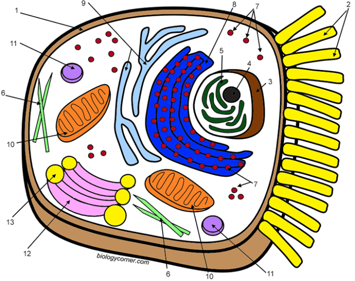
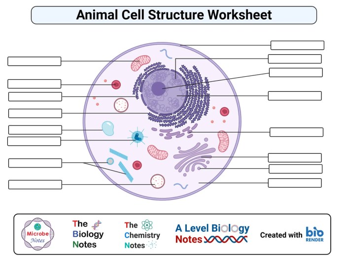
Animal cell coloring answer – The animal cell, a microcosm of life, reveals a breathtaking orchestration of intricate structures working in harmonious synergy. Each component plays a vital role, contributing to the cell’s overall vitality and perpetuation. Understanding these structures and their functions unveils a deeper appreciation for the profound complexity and elegance of biological systems. This exploration will illuminate the interconnectedness and purpose within this fundamental unit of life.
Cell Membrane Structure and Function
The cell membrane, a fluid mosaic of lipids and proteins, acts as the cell’s sentinel, regulating the passage of substances into and out of the cell. Its selectively permeable nature ensures the maintenance of a stable internal environment, crucial for cellular processes. The phospholipid bilayer, with its hydrophilic heads and hydrophobic tails, forms the structural backbone, while embedded proteins facilitate transport, signaling, and cell recognition.
This dynamic barrier is not merely a passive gatekeeper but an active participant in cellular communication and homeostasis.
The Nucleus: Orchestrator of Cellular Activities
The nucleus, often referred to as the cell’s control center, houses the genetic material, DNA. Within its double membrane envelope, DNA is organized into chromosomes, carrying the blueprint for the cell’s structure and function. The nucleus regulates gene expression, controlling which proteins are synthesized and when. This precise control ensures the cell’s proper development, maintenance, and response to its environment.
The nucleolus, a specialized region within the nucleus, is responsible for ribosome biogenesis, a critical step in protein synthesis.
Protein Synthesis: The Ribosome’s Vital Role, Animal cell coloring answer
Ribosomes, the protein factories of the cell, are the sites of protein synthesis. These complex molecular machines translate the genetic code from messenger RNA (mRNA) into the amino acid sequences that form proteins. Ribosomes can be found free-floating in the cytoplasm or attached to the endoplasmic reticulum. This process, involving transfer RNA (tRNA) carrying specific amino acids, is a fundamental process for building and maintaining cellular structures and functions.
The accuracy and efficiency of protein synthesis are paramount for cellular health and survival.
Endoplasmic Reticulum and Golgi Apparatus: Processing and Packaging
The endoplasmic reticulum (ER) and Golgi apparatus work in concert to process and package proteins and lipids. The rough ER, studded with ribosomes, synthesizes proteins destined for secretion or membrane insertion. The smooth ER, lacking ribosomes, synthesizes lipids and detoxifies harmful substances. The Golgi apparatus, a stack of flattened membrane-bound sacs, further modifies, sorts, and packages these molecules into vesicles for transport to their final destinations within or outside the cell.
This coordinated effort ensures the efficient distribution of cellular products.
Mitochondria: The Powerhouses of the Cell
Mitochondria, often called the “powerhouses” of the cell, are responsible for cellular respiration, generating ATP (adenosine triphosphate), the cell’s primary energy currency. These double-membraned organelles contain their own DNA and ribosomes, remnants of their endosymbiotic origins. The inner membrane’s intricate folds, called cristae, significantly increase the surface area for ATP production through oxidative phosphorylation. Mitochondria are essential for providing the energy needed for all cellular activities.
In contrast, chloroplasts, found only in plant cells, are responsible for photosynthesis, converting light energy into chemical energy.
Understanding the intricacies of an animal cell coloring answer requires a foundational knowledge of cellular structures. This understanding can be enhanced by exploring relatable visual aids, such as the charming illustrations found in farm animal coloring book pages , which, while not directly related to cellular biology, foster a similar appreciation for the detailed representation of living organisms.
Returning to the animal cell, accurate coloring helps solidify comprehension of the various organelles and their functions.
Animal Cell Organelles
| Name | Structure | Function | Location |
|---|---|---|---|
| Cell Membrane | Phospholipid bilayer with embedded proteins | Regulates passage of substances; cell signaling | Surrounds the cell |
| Nucleus | Double-membraned organelle containing DNA | Controls gene expression; houses genetic material | Center of the cell |
| Ribosomes | RNA and protein complexes | Protein synthesis | Free in cytoplasm or attached to ER |
| Endoplasmic Reticulum (ER) | Network of interconnected membranes | Protein and lipid synthesis; detoxification | Throughout cytoplasm |
| Golgi Apparatus | Stack of flattened membrane sacs | Processes, sorts, and packages proteins and lipids | Near the nucleus |
| Mitochondria | Double-membraned organelles with cristae | Cellular respiration; ATP production | Throughout cytoplasm |
Cell Coloring Techniques and Materials

The preparation of an animal cell slide for microscopic observation is a sacred act, a journey into the miniature universe that constitutes the very building blocks of life. Through careful preparation and staining, we unveil the intricate tapestry of cellular structures, revealing the divine artistry inherent in creation. This process allows us to glimpse the profound interconnectedness of all living things and appreciate the exquisite detail within each individual cell.
Preparing a slide for microscopic observation involves a delicate balance of precision and reverence. Each step, from sample collection to final staining, requires mindful attention to detail, reflecting our respect for the life we are studying. The resulting image, a vibrant tapestry of cellular structures, is a testament to the power of observation and the beauty inherent in the natural world.
Slide Preparation for Microscopic Observation
Preparing a suitable animal cell slide for microscopic examination is paramount for accurate observation. This process involves several crucial steps, each contributing to the final image’s clarity and accuracy. The following detailed steps ensure a successful preparation, revealing the cell’s intricate architecture.
- Sample Collection: Obtain a sample of animal cells, such as cheek cells (obtained by gently scraping the inside of the cheek with a sterile cotton swab), or a prepared blood smear. Handle the sample with care, recognizing its inherent value.
- Slide Preparation: Place a small drop of the cell sample onto a clean microscope slide. Ensure the drop is not too large, to avoid excessive spreading and overlapping cells.
- Smear Preparation (if necessary): For thicker samples like blood, gently spread the sample across the slide using another slide to create a thin, even layer. This allows for better light penetration during microscopy.
- Air Drying: Allow the slide to air dry completely. This prevents the formation of artifacts that can obscure cellular details.
- Heat Fixation (optional): Gently pass the slide through a flame a few times to heat-fix the cells. This process adheres the cells to the slide and enhances staining.
Appropriate Stains and Dyes
The selection of appropriate stains and dyes is crucial for visualizing specific cellular structures. Different stains possess unique affinities for different cellular components, allowing us to highlight specific features. This careful choice allows us to reveal the beauty and complexity hidden within the cell’s architecture.
- Methylene Blue: A general-purpose stain that readily stains the nucleus and cytoplasm, providing a basic overview of cellular morphology.
- Crystal Violet: Another general stain, similar to methylene blue, but often used in Gram staining techniques for bacteria. It stains the cell wall of bacteria.
- Eosin: A counterstain commonly used with hematoxylin; it stains the cytoplasm pink or red, contrasting with the blue-stained nucleus.
- Hematoxylin: A nuclear stain that intensely stains the cell nucleus a deep purple or blue, providing a clear delineation of this vital organelle.
Applying Stains for Optimal Visualization
The application of stains is a meditative process, requiring patience and a gentle touch. The goal is to achieve optimal staining without damaging the cellular structures or creating artifacts. Proper staining technique enhances the visualization of cellular components and reveals the intricate beauty of the cell.
The staining process typically involves flooding the prepared slide with the chosen stain for a specific duration, followed by rinsing with water or a buffer solution to remove excess stain. The optimal staining time varies depending on the stain and the type of cells being observed. Microscopic examination allows for the observation of the staining process and fine-tuning the technique.
Importance of Proper Slide Preparation
Proper slide preparation is the foundation of accurate microscopic observation. Without careful preparation, artifacts and distortions can obscure cellular structures, leading to inaccurate interpretations. Careful preparation ensures that the observed structures accurately reflect the cell’s true morphology and internal organization. It is a crucial step in gaining insight into the cellular world.
Careful preparation allows for the clear visualization of the cell’s intricate structure, revealing the wonders of cellular organization and the underlying mechanisms of life. The precision and care invested in slide preparation reflect our respect for the living world and our commitment to uncovering its mysteries.
Illustrated Guide to Staining Animal Cells
[Imagine a series of three simple drawings. Drawing 1: A simple circle representing a cell with a darker circle inside representing the nucleus. Drawing 2: The same cell, but the nucleus is now a vibrant blue, indicating staining with hematoxylin. Drawing 3: The same cell, but the cytoplasm is now a light pink, indicating the application of eosin as a counterstain.
The nucleus remains blue.]
Drawing 1: The unstained animal cell is shown, with a visible nucleus but lacking distinct features. This stage emphasizes the importance of staining to visualize internal structures.
Drawing 2: The application of hematoxylin intensely stains the nucleus blue, clearly defining its boundaries and structure. This highlights the nucleus’s importance as the cell’s control center.
Drawing 3: The addition of eosin as a counterstain gently colors the cytoplasm a light pink, providing a stark contrast to the blue nucleus and revealing the cytoplasm’s texture and composition. This allows for the observation of both the nucleus and the cytoplasm.
Interpreting Colored Animal Cell Slides: Animal Cell Coloring Answer

Gazing upon a stained animal cell slide is akin to peering into a microcosm of life, a vibrant tapestry woven from the threads of cellular machinery. The colors, shapes, and relative sizes of the organelles revealed through staining offer a window into the cell’s dynamic processes, its very essence. Understanding these visual cues unlocks a deeper appreciation for the intricate workings of life itself.The vibrant hues of a stained animal cell are not merely aesthetic; they are signposts guiding us to the hidden wonders within.
Each color represents a specific organelle, its intensity reflecting the concentration of the stained substance. By meticulously analyzing these chromatic indicators, we can unravel the cell’s functional narrative.
Key Organelles Visible in Stained Animal Cell Slides
Careful observation reveals several key organelles within a stained animal cell. The nucleus, often a prominent, darkly stained sphere, houses the cell’s genetic material. The cytoplasm, the jelly-like substance filling the cell, may appear differently stained depending on the technique used. Mitochondria, the powerhouses of the cell, often appear as small, rod-shaped structures, staining differently depending on the dye used.
The endoplasmic reticulum, a network of membranes involved in protein synthesis and lipid metabolism, may appear as a faintly stained, interconnected network. Finally, the Golgi apparatus, responsible for modifying, sorting, and packaging proteins, may appear as a stack of flattened sacs, often stained differently from the surrounding cytoplasm.
Differentiating Organelles Based on Color and Shape
Distinguishing between organelles relies on a combination of their characteristic staining patterns and morphologies. For instance, the nucleus, typically large and round, stands out due to its intense staining with DNA-specific dyes. Mitochondria, smaller and rod-shaped, often exhibit a different staining intensity compared to the nucleus or cytoplasm, depending on the dye’s affinity for their components. The endoplasmic reticulum’s network-like structure and faint staining contrast with the more distinctly stained nucleus and mitochondria.
The Golgi apparatus’s stacked structure also provides a visual cue for identification. Variations in staining intensity may arise from differences in the dye used, the fixation process, and the cell’s physiological state.
Significance of Observing Specific Organelles and Staining Patterns
The observation of specific organelles and their staining patterns is crucial for understanding cellular function and health. Variations in staining intensity or morphology can indicate cellular stress, disease, or other physiological changes. For example, an unusually pale staining of mitochondria might suggest mitochondrial dysfunction, while an abnormally large or irregularly shaped nucleus could point to genetic abnormalities. Such observations are essential in various fields, including medical diagnostics, biological research, and environmental monitoring.
Comparative Analysis of Different Staining Techniques
Different staining techniques highlight different cellular components, providing a multifaceted view of the cell’s structure and function. The choice of staining technique depends on the specific organelles of interest and the research question. Here’s a comparison of a few common techniques:
Staining Technique
Primary Stain
Organelles Highlighted
Visual Characteristics


Hematoxylin and Eosin (H&E)
Hematoxylin (basic dye), Eosin (acidic dye)
Nucleus, cytoplasm, connective tissue
Nucleus: dark purple/blue; Cytoplasm: pink/red; Connective tissue: varying shades of pink


Wright-Giemsa
Methylene blue, eosin, azure B
Blood cells, microorganisms
Differentiation of various blood cell types based on color and morphology


Sudan Black B
Sudan Black B
Lipids
Lipids appear black against a lighter background


Periodic Acid-Schiff (PAS)
Periodic acid, Schiff's reagent
Carbohydrates
Carbohydrates stain magenta/pink
Common Errors and Troubleshooting in Cell Coloring

The journey of revealing the intricate beauty of the animal cell through staining is a path of precision and mindful observation. Like a skilled artisan carefully applying color to a masterpiece, the cell biologist must approach the staining process with attentiveness and understanding. Imperfect technique can obscure the very structures we seek to illuminate, leading to misinterpretations and hindering our understanding of this fundamental unit of life.
Therefore, recognizing and addressing common errors is paramount to achieving accurate and insightful results.Uneven Staining and Poor Cell Visualization: These are frequent challenges encountered during the staining process. Uneven staining can arise from inadequate mixing of the dye solution, insufficient staining time, or the presence of interfering substances within the sample. Poor cell visualization, on the other hand, often stems from the use of an inappropriate dye concentration or a failure to properly prepare the cell sample.
The consequences of these errors range from difficulty in identifying cellular components to complete misinterpretation of cell morphology and structure.
Causes and Solutions for Uneven Staining
Uneven staining, a common pitfall, often results from inadequate mixing of the stain, leading to variations in dye concentration across the slide. This uneven distribution can create areas of over-staining, obscuring detail, and areas of under-staining, rendering structures invisible. Another cause is insufficient contact time between the cells and the stain. Finally, the presence of debris or interfering substances within the sample can prevent the dye from penetrating the cells uniformly.
Solutions include ensuring thorough mixing of the stain, optimizing staining time through experimentation, and meticulously cleaning the cell preparation to remove any interfering materials.
Importance of Controls in Staining
The use of appropriate controls is essential in validating the staining procedure and ensuring the accuracy of the results. Positive controls, using cells known to stain effectively with the chosen dye, confirm that the staining solution is functioning correctly. Negative controls, employing cells or a blank slide without the stain, establish a baseline and help identify non-specific staining or background interference.
The inclusion of controls acts as a crucial safeguard, preventing misinterpretations arising from technical errors or limitations of the staining technique.
Impact of Staining Time and Dye Concentration
Variations in staining time and dye concentration significantly impact the quality and interpretability of the stained cell preparation. Prolonged staining can lead to over-staining, obscuring fine details and potentially damaging cellular structures. Conversely, insufficient staining time may result in under-staining, making it difficult to visualize the targeted structures. Similarly, using an excessively high dye concentration can result in over-staining and background staining, while a concentration that is too low can yield weak or indistinct staining.
Careful optimization of both parameters is crucial to achieve optimal staining results.
Troubleshooting Guide
The following guide provides solutions to common problems encountered during animal cell staining. Remember, patience and careful observation are key to success.
“Problem: Uneven staining. Solution: Ensure thorough mixing of the stain, optimize staining time, and meticulously clean the cell preparation.”
“Problem: Poor cell visualization. Solution: Adjust dye concentration, optimize staining time, and ensure proper sample preparation.”
“Problem: Excessive background staining. Solution: Filter the staining solution, reduce staining time, or use a lower dye concentration.”
“Problem: Cells appear shrunken or distorted. Solution: Use a gentler fixation method, or reduce the duration of fixation.”
“Problem: No staining observed. Solution: Verify dye viability, check staining time and technique, and ensure proper sample preparation.”
