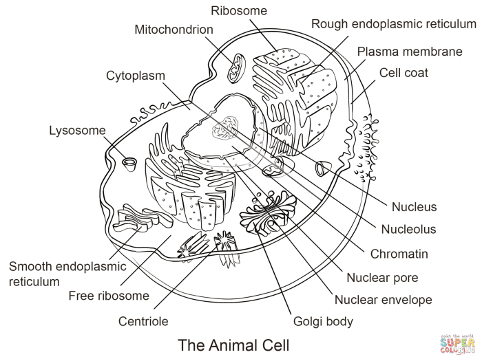
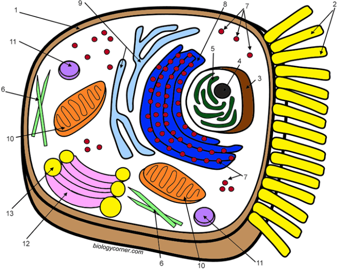
Creating Educational Content for the Coloring Page

Animal cell coloring printable – This coloring page provides a fun and engaging way to learn about the fascinating world of animal cells. By coloring the different parts of the cell, children can visualize their structure and begin to understand their vital functions. This section Artikels educational content suitable for various age groups, explains the importance of cell structure and function, and suggests ways to use the coloring page as a learning tool.
Animal Cell Facts for Different Age Groups
Understanding animal cells is crucial for grasping fundamental biological concepts. The following facts are categorized by age group to ensure appropriate complexity and engagement.
- Elementary School (Ages 6-10): Animal cells are like tiny building blocks that make up all animals, including you! They have a nucleus, which is like the brain of the cell, and a cell membrane, which is like the skin, protecting everything inside. The cytoplasm is the jelly-like substance filling the cell, and it helps keep everything in place. Think of the cell as a tiny, busy city with different parts working together.
- Middle School (Ages 11-14): Animal cells are eukaryotic cells, meaning they have a membrane-bound nucleus containing their genetic material (DNA). They also contain various organelles, including mitochondria (the powerhouses of the cell), ribosomes (protein factories), and the endoplasmic reticulum (a network involved in protein and lipid synthesis). The Golgi apparatus processes and packages proteins, while lysosomes break down waste. Understanding these organelles helps explain how cells carry out essential life processes.
The Importance of Cell Structure and Function in Maintaining Life
The structure of an animal cell is directly related to its function. Each organelle plays a specific role in maintaining the cell’s health and enabling it to perform its tasks. For example, the mitochondria generate energy through cellular respiration, providing the power needed for all cellular processes. The nucleus houses the genetic information that dictates the cell’s activities and ensures proper cell division.
Disruptions in the structure or function of any organelle can lead to cellular dysfunction and potentially disease. The coordinated activity of all organelles is essential for maintaining the life of the organism.
Using the Coloring Page as a Learning Tool
The coloring page can be a valuable tool for learning about animal cells. Students can:
- Label the parts: After coloring, encourage students to label each organelle on their drawing using a textbook or online resource as a guide.
- Create a model: The coloring page can serve as a template for creating a three-dimensional model of an animal cell using various materials like clay, playdough, or even recycled materials.
- Research and report: Assign students to research a specific organelle and create a short report on its function and importance within the cell.
Animal Cell Quiz
This quiz can be used to assess understanding after completing the coloring page.
Animal cell coloring printables are a great educational tool for understanding cell structures. Interestingly, the vibrant colors often used in these printables can sometimes be derived from sources like insects, as you might discover when researching animal based food coloring. This connection highlights the surprising origins of some common pigments, making the simple act of coloring an animal cell a more engaging learning experience.
- What is the control center of the animal cell?
- What organelle produces energy for the cell?
- What is the jelly-like substance that fills the cell?
- What is the function of the cell membrane?
- Name one other organelle found in animal cells (besides those mentioned above).
Illustrating Animal Cell Components

This section provides detailed descriptions of the key components of an animal cell, focusing on their visual representation for a coloring page. Accurate depiction is crucial for understanding their functions and relationships within the cell. The descriptions below aim to guide the creation of a clear and informative illustration.
Nucleus
The nucleus, the cell’s control center, should be depicted as a large, round or oval structure, generally centrally located. Its boundary, the nuclear envelope, is a double membrane with numerous pores, which should be shown as small dots or openings scattered across its surface. The interior, the nucleoplasm, can be slightly shaded differently to distinguish it from the cytoplasm.
A prominent, darker area within the nucleus represents the nucleolus, responsible for ribosome production. Consider adding a slightly textured appearance to the nucleoplasm to suggest the presence of chromatin (DNA).
Cytoplasm
The cytoplasm is the jelly-like substance filling the cell, excluding the nucleus. It should be represented as a light-colored background filling the space between the cell membrane and the organelles. Avoid making it uniformly colored; subtle shading or texture can suggest its complex composition. The cytoplasm houses all the other organelles and facilitates cellular processes.
Mitochondria
Mitochondria, the powerhouses of the cell, should be depicted as elongated or oval structures with a folded inner membrane. These folds, called cristae, significantly increase the surface area for energy production and should be visible as internal ridges or lines within each mitochondrion. They should be shown scattered throughout the cytoplasm. Their double membrane structure – an outer and an inner membrane – can be suggested by subtle shading differences.
Ribosomes, Animal cell coloring printable
Ribosomes, the protein synthesis factories, are extremely small and should be depicted as tiny dots scattered throughout the cytoplasm, sometimes attached to the endoplasmic reticulum. Their small size necessitates a stylistic representation rather than a detailed anatomical rendering. They may be shown as clusters in some areas, reflecting their aggregation during protein production.
Endoplasmic Reticulum
The endoplasmic reticulum (ER) is a network of interconnected membranes. The rough ER, studded with ribosomes, should be illustrated as a series of interconnected flattened sacs or cisternae with small dots (ribosomes) attached to its surface. The smooth ER, lacking ribosomes, should be shown as a network of interconnected tubules, appearing smoother and less studded than the rough ER.
These two forms should be shown interacting, demonstrating their interconnected nature.
Golgi Apparatus
The Golgi apparatus, or Golgi body, should be depicted as a stack of flattened, membrane-bound sacs or cisternae. These sacs should be slightly curved and arranged in a somewhat layered fashion. Small vesicles, representing the transport of proteins, can be shown budding off from the edges of the Golgi stacks.
Lysosomes
Lysosomes are small, membrane-bound organelles containing digestive enzymes. They should be shown as small, oval or spherical structures scattered throughout the cytoplasm. A slightly darker shading inside could subtly indicate the presence of digestive enzymes.
Vacuoles
Vacuoles are membrane-bound sacs used for storage. In animal cells, they are generally smaller and more numerous than in plant cells. They should be represented as small, irregularly shaped, clear vesicles within the cytoplasm. They can be shown containing different shades to suggest various stored substances.
Question Bank: Animal Cell Coloring Printable
What materials are needed to use the animal cell coloring printable?
Colored pencils, crayons, markers, or paint are all suitable. Paper is, of course, essential.
Can this printable be used for different age groups?
Yes, the design can be adapted to suit different age groups. Simpler versions can be created for younger children, while more detailed versions can be used for older students.
Where can I find additional information about animal cell structures?
Numerous online resources and textbooks provide detailed information about animal cell structures and functions. A simple online search will yield many results.
How can I assess student understanding after using the coloring page?
Include a short quiz or worksheet with questions about the cell organelles and their functions. Observe student work and participation during the activity.
