Coloring Schemes for Animal Cell Organelles: Animal Cell Coloring Different Colour

Animal cell coloring different colour – Assigning distinct colors to different organelles in an animal cell diagram is crucial for enhancing understanding and memorability. A well-chosen color scheme improves the clarity of the diagram, making it easier to identify and differentiate various cellular components and their functions. Effective color choices can also help students visualize the spatial relationships between organelles and their roles in complex cellular processes.Color schemes for animal cell diagrams should prioritize clarity and educational effectiveness.
Illustrating the diverse components of an animal cell through color is a fun and educational activity. The varied organelles, each with a specific function, lend themselves well to creative expression, much like the playful designs found in a animal cartwheel coloring sheet. Similarly, choosing distinct colors for the nucleus, mitochondria, and other cellular structures enhances understanding and visual appeal of the animal cell’s intricate internal workings.
The selection of colors should be guided by the need to visually distinguish organelles, facilitating comprehension of their individual roles and their interactions within the cell. Furthermore, color choices should be mindful of potential color blindness issues, ensuring accessibility for all learners.
Comparison of Three Color Schemes
Three distinct color schemes can be compared to highlight their relative strengths and weaknesses in representing an animal cell. These schemes differ in their approach to color assignment, focusing on either functional grouping, structural similarity, or a more arbitrary but visually distinct approach.
- Scheme 1: Function-Based Coloring: This scheme groups organelles based on their primary functions. For example, all organelles involved in protein synthesis (ribosomes, rough endoplasmic reticulum, Golgi apparatus) might be shaded in variations of blue, while organelles involved in energy production (mitochondria) could be in shades of red. This approach aids in understanding the interconnectedness of cellular processes. However, it might lead to visually similar organelles being grouped together, even if they are structurally different.
- Scheme 2: Structure-Based Coloring: This approach assigns colors based on the structural characteristics of the organelles. Membrane-bound organelles might be depicted in shades of green, while non-membrane-bound organelles could be represented in shades of yellow. This method helps students understand the different levels of organization within the cell. However, it may not be as effective in highlighting functional relationships between organelles.
- Scheme 3: Visually Distinct Coloring: This approach prioritizes visual contrast and memorability. Each organelle is assigned a distinct, easily recognizable color, regardless of its function or structure. This scheme may be easier for initial identification, but it might not be as effective in conveying functional relationships or structural similarities.
Examples of Effective Color Palettes
Several color palettes can effectively represent the functions of different organelles. For instance, a vibrant green could represent the chloroplasts (although absent in animal cells, this is included for illustrative purposes of functional color assignment and can be substituted with a similarly bright color for another organelle), highlighting their role in photosynthesis (if applicable to a plant cell). A deep red could represent mitochondria, reflecting their role in energy production.
A light blue could be used for the endoplasmic reticulum, highlighting its role in transport and protein synthesis. Using analogous colors (colors next to each other on the color wheel) for organelles with related functions can improve visual coherence.
A Color Scheme Emphasizing Interconnectedness
To emphasize the interconnectedness of organelles, a color scheme can utilize a gradient or transition between colors. For example, the nucleus (control center) could be a dark purple, transitioning to lighter shades of purple for the endoplasmic reticulum and Golgi apparatus (involved in protein synthesis based on nuclear instructions). Mitochondria could be a distinct color (e.g., orange) but its proximity to other organelles involved in energy utilization could be subtly highlighted through color choice and placement.
This scheme helps visually represent the flow of information and materials within the cell. The lysosomes could be a contrasting color (e.g., deep blue) to highlight their role in waste breakdown, a process separate but connected to the overall cellular function.
Illustrative Examples of Animal Cell Coloring

A visually appealing and informative animal cell diagram relies heavily on effective color choices. The colors selected should not only be aesthetically pleasing but also enhance understanding of the organelles’ functions and relationships within the cell. A well-designed color scheme aids in quickly identifying and differentiating various components, ultimately improving comprehension.
Cell Membrane Representation, Animal cell coloring different colour
The cell membrane, a crucial structure regulating what enters and exits the cell, can be depicted using a gradient of dark blue to light blue. The darker shade represents the hydrophobic tails of the phospholipid bilayer, while the lighter blue signifies the hydrophilic heads. This gradient visually emphasizes the membrane’s dual nature and its role as a selective barrier.
A thin, lighter grey line could be added to represent the embedded proteins, further highlighting the membrane’s dynamic and complex structure. The overall effect is a visually engaging representation of the membrane’s selective permeability.
Golgi Apparatus Illustration
The Golgi apparatus, responsible for modifying, sorting, and packaging proteins, can be illustrated using a series of stacked, flattened sacs. These sacs, or cisternae, could be depicted in varying shades of orange, progressing from a lighter orange at the cis face (receiving end) to a deeper, richer orange at the trans face (shipping end). This color gradient represents the progression of protein modification as they move through the Golgi.
Small, dark brown dots could represent the proteins undergoing modification and transport within the cisternae. The shading could also emphasize the curvature of the sacs, adding to the three-dimensional effect.
Endoplasmic Reticulum Differentiation
The rough endoplasmic reticulum (RER) and smooth endoplasmic reticulum (SER) can be easily differentiated through color choices. The RER, studded with ribosomes, can be represented in a light teal color, with small dark purple dots representing the ribosomes. This highlights its role in protein synthesis. In contrast, the SER, involved in lipid synthesis and detoxification, can be depicted in a lighter, pastel yellow.
The difference in color and texture (due to the presence or absence of ribosomes) clearly distinguishes the two types of ER, emphasizing their distinct functions within the cell.
Animal Cell Diagram: A Visual Description
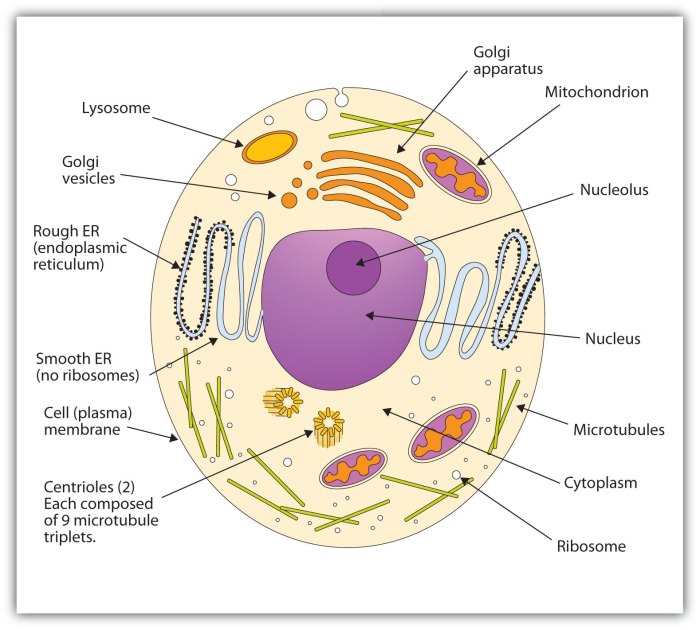
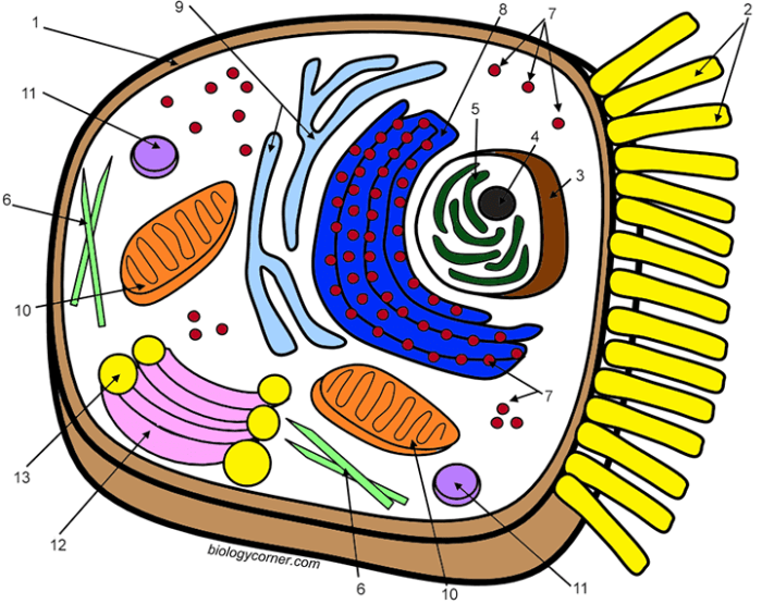
In this example, the cell membrane is a gradient of deep teal to light teal. The nucleus is a deep purple, with a lighter purple nucleolus. The rough endoplasmic reticulum is a light green dotted with dark green ribosomes. The smooth endoplasmic reticulum is a pale yellow. The Golgi apparatus is a series of flattened sacs, transitioning from light orange to a deeper burnt orange.
Mitochondria are depicted in a vibrant red, emphasizing their energy-producing role. Lysosomes are shown as small, dark magenta spheres. The cytoskeleton is represented by a thin, light grey network throughout the cell, providing structural support. The cytoplasm is a light beige, providing a neutral background that allows the organelles to stand out clearly. These color choices offer a clear and informative visual representation of the animal cell’s internal organization and the functions of its various components.
The use of color gradients and shading adds depth and visual interest, while the distinct colors allow for easy identification of each organelle.
Questions Often Asked
What are the best tools for digital animal cell coloring?
Digital art programs like Adobe Photoshop, Procreate, or even free online tools like Google Drawings offer excellent capabilities for creating detailed and colorful animal cell diagrams.
How can I make my animal cell coloring project more visually appealing?
Incorporate shading and texture to add depth and realism. Experiment with different color palettes to find one that enhances the clarity and visual appeal of your diagram. Consider adding labels for key organelles to improve understanding.
Are there any specific color combinations to avoid when coloring animal cells?
Avoid using colors that are too similar, as this can make it difficult to distinguish between different organelles. Also, avoid overly bright or jarring color combinations that could detract from the educational purpose.
